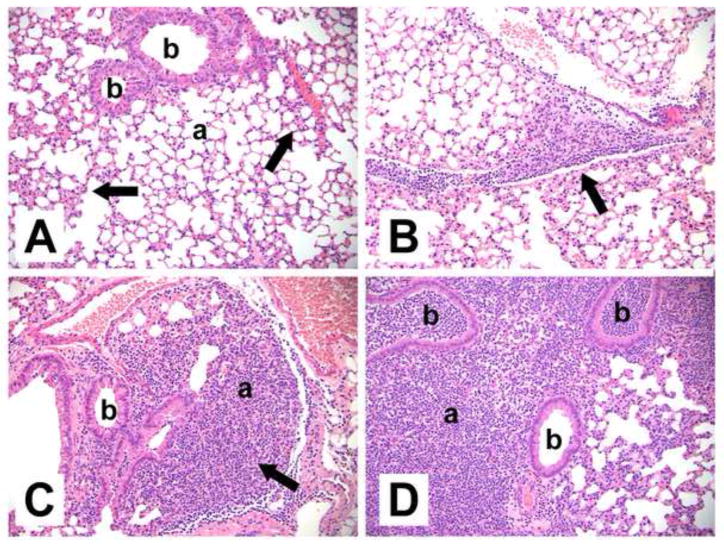
Fig. 3. Representative histopathological changes in the lungs of S. pneumoniae-infected wild type (WT) and EP2 knockout (KO) mice 24 h after inoculation.
Abbreviations: b: bronchiole, a: alveoli. (A) Infected WT mouse without significant inflammation (summary score 0.5). The majority of alveolar airspaces are clear. There are small areas of increased interstitial macrophagic cellularity (arrows). (B) Infected WT mouse with mild, focal neutrophilic inflammation (summary score 1.0), adjacent to a pulmonary blood vessel and along interlobular septae (arrow). (C) Infected EP2−/− mouse with mild-moderate inflammation (summary score 1.5) focally obscuring the alveoli (arrow). (D) Infected EP2−/− mouse with focally intense inflammation (summary score 7.5) consisting of neutrophils extensively filling alveoli and bronchioles, pleural involvement (not shown), and increased interstitial cellularity. Hematoxylin and eosin. Original magnifications x200.

