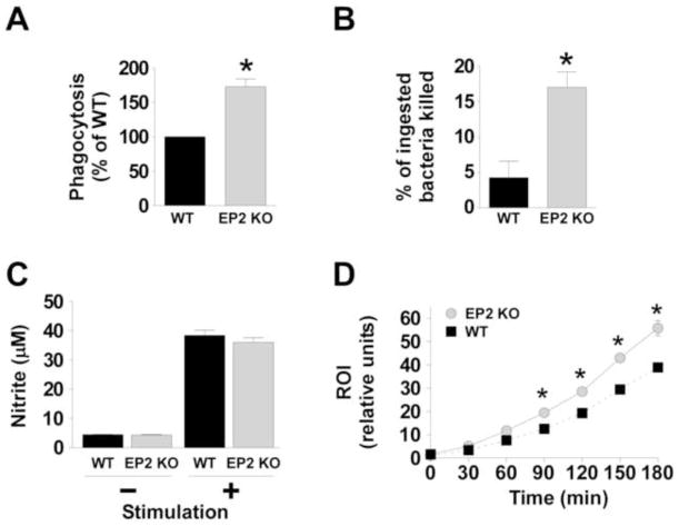
Fig. 5. Enhanced in vitro alveolar macrophage defense functions in EP2 knockout (KO) mice compared with wild type (WT) alveolar macrophages.
Alveolar macrophages were obtained from uninfected WT and EP2 KO mice as described in the Materials and Methods section. (A) Cells were inoculated with heat-inactivated, FITC-labeled S. pneumoniae (multiple of infection 150 bacteria per macrophage) and phagocytosis was measured after 3 hrs. *P < 0.05 by Student t-test. Data represent the mean ± SEM of a minimum of 3 independent experiments performed in octuplet. (B) The survival of internalized S. pneumoniae was determined as noted in the Materials and Methods section. *P < 0.05 by Student t-test. Data represent the mean ± SEM of a minimum of 3 independent experiments performed in triplicate. (C) The capacity for alveolar macrophages to generate nitric oxide (measured as nitrite) was assessed for WT and EP2 KO cells following stimulation with or without 10 μg/ml of lipoteichoic acid and 10 ng/ml IFN-γ for 24h. (D) Alveolar macrophages from WT mice (black squares and dotted line) or EP2 KO mice (grey circles and solid line) were cultured with 2′,7′-dichlorodihydrofluorescein diacetate (H2DCF) for 1h then stimulated with heat-killed S. pneumoniae using a multiplicity of infection of 50:1. Reactive oxygen intermediate (ROI) production was assessed fluorometrically and expressed as relative fluorescence units. The data represent the mean of 3 experiments completed in quadruplicate for each time point. *P < 0.05 by Student t-test.

