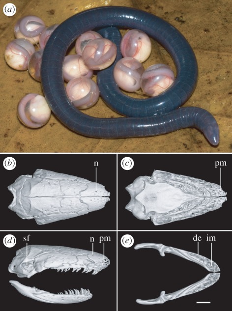
Figure 3.
Morphology of Chikilidae fam. nov. (a) Chikila fulleri in life, brooding egg clutch (in captivity). Scale bar, 10 mm. (b–e) Volume reconstruction of high-resolution X-ray computed tomography data showing cranium and mandibles of C. fulleri. (b) Cranium in dorsal view. (c) Cranium in palatal view. (d) Cranium and mandible in right lateral view. (e) Mandibles in dorsal view. Scale bar (b,c), 1 mm. n, nasal; pm, premaxilla; sf, stapedial foramen; im, inner mandibular (i.e. ‘splenial’) tooth row; de, dentary tooth row. See the electronic supplementary material, figure S2 for more detailed labelling.

