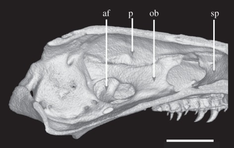
Figure 4.
Volume reconstruction of high-resolution X-ray computed tomography data showing sagittal section through braincase of Chikila fulleri (SDB 1304). The left internal wall of the braincase is seen from the midline, showing the single antotic foramen, and exclusion of the frontal from the roof of the braincase posterior to the sphenethmoid (formed by the parietal). af, antotic foramen; ob, os basale; p, parietal; sp, sphenethmoid. Scale bar, 1 mm.

