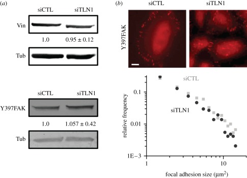
Figure 3.
Effect of talin-1 depletion on focal adhesion composition and size distribution. (a) Western blots of vinculin and pY397FAK in siCTL and siTLN1 cells revealed no significant alteration in the expression levels of these two proteins after treatment with RNAi for 3 days. Tubulin (Tub) levels are used as loading controls. As in figure 1, the numbers below the blots indicate densitometric band intensities (mean ± s.d. over n ≥ 3 blots) normalized to the corresponding siCTL value. (b) Localization of pY397FAK at focal adhesions in siCTL and siTLN1 cells. Scale bar, 20 µm. The plot depicts log–log histograms of focal adhesion areas for siCTL (black circles) and siTLN1 (grey squares), as determined from pY397FAK immunofluorescence images (see §2 for details on data acquisition and processing). The strong overlap between the two histograms implies similar focal adhesion size distributions for siCTL and siTLN1 cells. (Online version in colour.)

