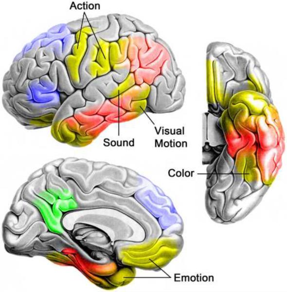
Figure 4. A neuroanatomical model of semantic processing.
A model of semantic processing in the human brain is shown, based on a broad range of pathological and functional neuroimaging data. Modality-specific sensory, action, and emotion systems (yellow regions) provide experiential input to high-level temporal and inferior parietal convergence zones (red regions) that store increasingly abstract representations of entity and event knowledge. Dorsomedial and inferior prefrontal cortices (blue regions) control the goal-directed activation and selection of the information stored in temporoparietal cortices. The posterior cingulate gyrus and adjacent precuneus (green region) may function as an interface between the semantic network and the hippocampal memory system, helping to encode meaningful events into episodic memory. A similar, somewhat less extensive semantic network exists in the right hemisphere, although the functional and anatomical differences between left and right brain semantic systems are still unclear.

