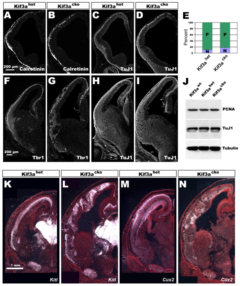
Figure 2. Neurogenesis occurs normally in Kif3acko cortices.
(A–D) Immunostaining of E11.5 coronal sections of Kif3ahet (A and C) and Kif3acko (B and D) brains. Calretinin labels Cajal Retzius cells and TuJ1 is a pan-neuronal marker. (E) Quit fraction analysis of E12.5 cortices. About 7% more neurons are produced in Kif3acko than Kif3ahet cortices in a 24-hour period. (F–I) Immunostaining of E13.5 coronal sections of Kif3ahet (F and H) and Kif3acko (G and I) brains. Tbr1 labels preplate and layer 6 neurons. (K–N) In situ hybridizations of E18.5 coronal sections showing that fate specification is normal in Kif3acko brains. Kitl marks deep-layer neurons and Cux2 labels superficial neurons.

