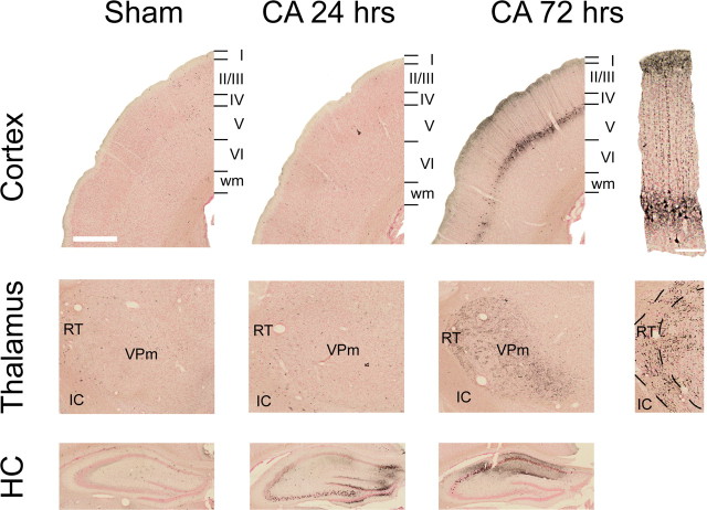
Figure 8.
Neurodegeneration in the rat brain after a 9 min CA or sham insult. Degenerating neurons and neurites were visualized with Amino Cupric silver stain. Top row: in the cortex, degeneration of Layer V pyramidal neurons and their apical dendrites is apparent at 72 h. Middle row: in the thalamus, neurites in VPm and in RT also degenerate 72 h after CA. Bottom row: in the hippocampus, neuronal degeneration in CA2, CA3, and the dentate gyrus peaks at 24 h after CA, whereas degeneration in CA1 becomes more pronounced 72 h after CA. Scale bars: 500 μm (low-power images); 100 μm (high-power images).

