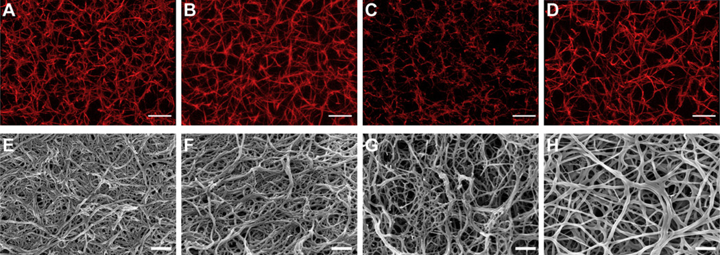
Fig. 2.
Confocal and scanning electron microscopy: Representative confocal micrograph projections (10 µm z-stacks; A–D) and SEM images (E–H) from no peptide control (A and E), control peptide conjugate, GPSPFPAC-PEG (B and F), knob ‘A’ conjugate, GPRPFPAC-PEG (C and G), and knob ‘B’ conjugate, AHRPYAAC-PEG (D and H). Confocal: Objective = 63×, Scale bar = 10 µm. SEM: Magnification = 50,000×, Voltage = 3.0 kV, Scale bar = 500 nm.

