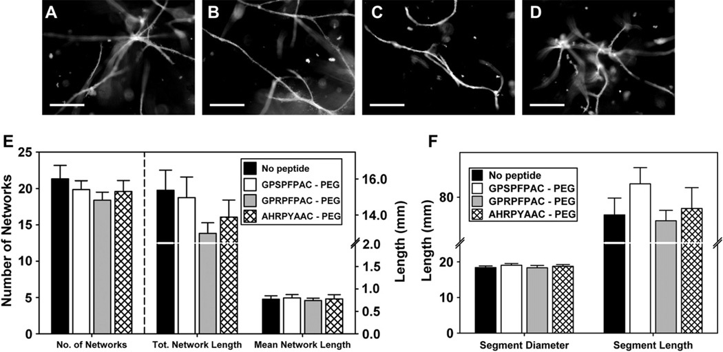
Fig. 7.
Angiogenic sprouting from microvessel fragments: Representative fluorescence micrographs of constructs at day 10 from no peptide control (A), control peptide (GPSPFPAC-PEG; B), knob ‘A’ conjugate (GPRPFPAC-PEG; C), and knob ‘B’ conjugate (AHRPYAAC; D). Scale bar = 200 µm. Comparison of network characteristics across the substrates (E), Comparison of segment characteristics across the substrates (F). Note that multiple connected segments are contained within a network. No significant difference was observed between groups.

