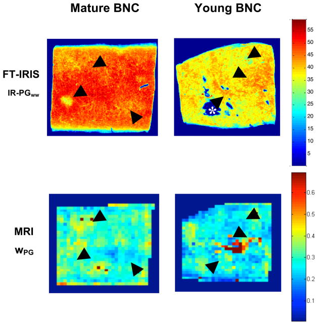
Figure 1.
Representative FT-IRIS normalized PG maps (IR-PGww) and MRI-derived PG-bound water maps (wPG) from young and mature BNC. Normalization, as described in the text, allows for a more direct comparison between modalities since FT-IRIS measurements are made on histologically prepared tissue sections while MRI measurements are made on intact hydrated samples. Black arrows highlight corresponding regions. An example of the voids caused by histological sectioning is indicated by the white asterisk (*).

