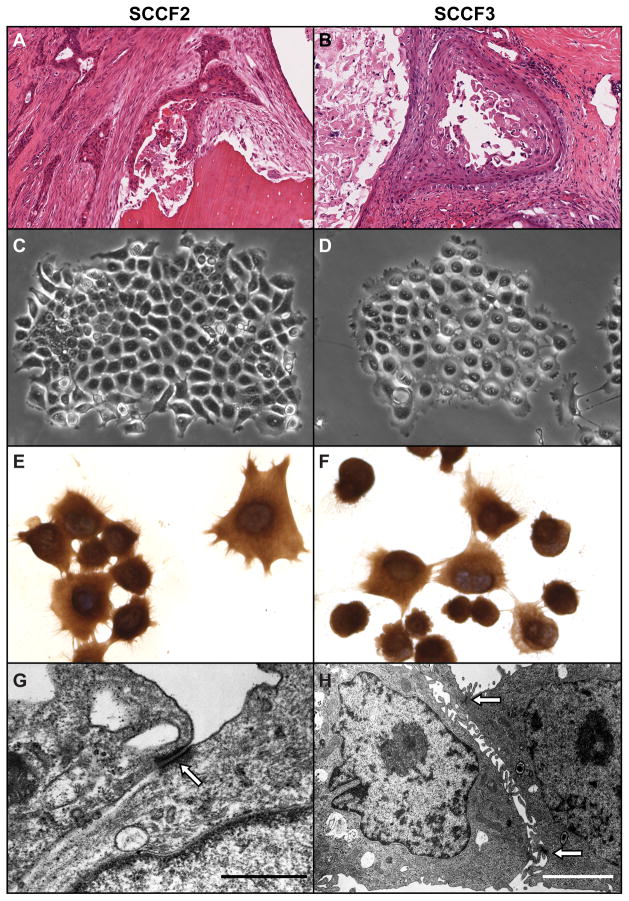
Figure 1. SCCF2 and SCCF2 morphology.
SCCF2 and SCCF3 cell lines were derived from spontaneous feline OSCC cancers. A. SCCF2 originated from a bone invasive maxillary OSCC . OSCC cells formed thin cords occasionally surrounding foci of keratin and sloughed tumor cells, within dense fibrous stroma and adjacent to a fragment of partially resorbed, necrotic, lamellar bone (Hematoxylin and eosin (HE)). B. SCCF3 was derived from an invasive lingual OSCC. The tumor was composed of large cystic spaces lined by neoplastic squamous epithelium and contained large quantities of keratin and necrotic tumor cells. SCCF2 (C) and SCCF3 (D) cells both grew in vitro as colonies of adherent cells with typical round to polygonal cell morphology. SCCF2 (E) and SCCF3 (F) cells grown on glass slides were cytokeratin positive (Pancytokeratin immunocytochemistry, DAB and hematoxylin counterstain). SCCF2 (G) and SCCF3 (H) cells were grown on glass and were examined using transmission electron microscopy. Both cell types demonstrated desmosomes characteristic (open arrow) of epithelial cells (Scale bars: G, 0.7 μM; H, 2.5 μM).

