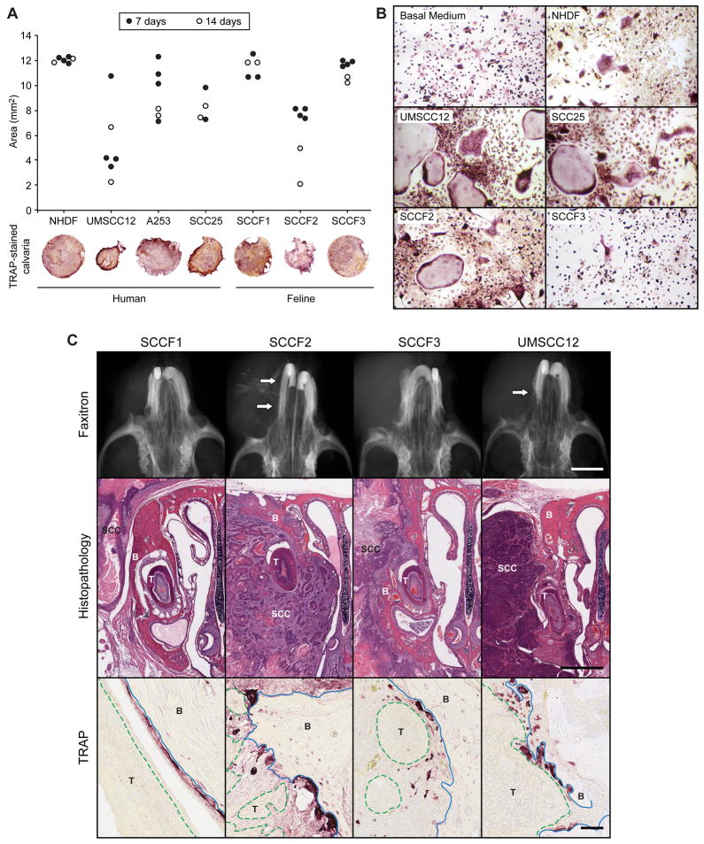
Figure 2. Feline and human OSCC cells induced in vitro and in vivo osteoclastic bone resorption.
A. Murine calvarial bone discs (4 mm diameter) were co-cultured with OSCC cells for 7 (black circles) or 14 (white circles) days. Calvaria were fixed, stained for TRAP activity, photographed and evaluated for changes in bone area using histomorphometry. SCCF2 (feline) and UMSCC12 (human) cells stimulated the most bone resorption. B. Murine BMSCs were cultured in CM from human and feline OSCC cells. UMSCC12, SCC25 and SCCF2 cells stimulated the formation of numerous, large osteoclasts. C. In vivo, minimal to no evidence of bone resorption was radiographically evident in SCCF1 and SCCF3-tumor-bearing mice. Moderate to marked reactive bone resorption (white arrows) was observed in the SCCF2Luc and UMSCC12Luc-bearing mice. SCCF1Luc xenografts were rarely associated with bone resorption despite close proximity of tumor to bone. SCCF2Luc xenografts were characterized by marked bone loss (xenograft indicated by ‘SCC’, tooth by ‘T’ and bone by ‘B’) and invasion with the formation of numerous large, TRAP-positive osteoclasts. Osteoclasts are red-stained cells on the surface of bone (blue solid line) adjacent to OSCC xenografts (green dashed line, labeled ‘T’). Less bone invasion was observed in the SCCF3Luc xenografts; however, osteoclastic bone resorption was occasionally observed. UMSCC12Luc frequently invaded bone and stimulated osteoclastic bone resorption. Scale bars: Faxitron: 1 mm; Histopathology: 1 mm; TRAP: 50 μm.

