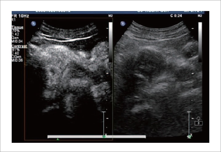
Figure 3.

CE US revealed a hypoechoic mass of 4.7 cm×3.4 cm in the pancreatic head, with blurred delineated margins. In the perfusion image phase, harmonic imaging demonstrated enhancement of the mass in its arterial phase using agent detection imaging mode approximately synchronizing with the rest of the pancreas.
