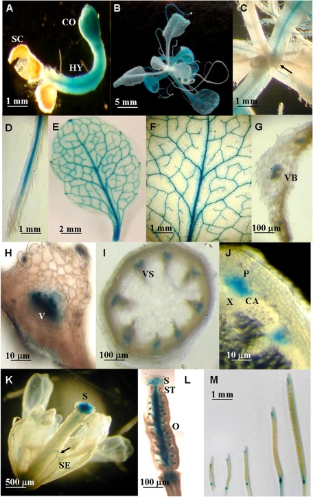
Fig. 2.
Spatial and temporal expression patterns of ACBP3pro::GUS fusions. Histochemical GUS staining shows expression of GUS from the ACBP3 5′-flanking region in a 3-day-old seedling (A); a 3-week-old seedling (B and C), with the arrow in C showing newly produced leaves; root (D); 32-day-old rosette leaf with the major and side veins (E and F); horizontal section of leaf (G and H); stem (I and J); fully opened flower, with the arrow showing the sepal (K); hand-section of a pistil showing the expression of ACBP3pro::GUS in stigma, style, and ovary (L); siliques from a 40-day-old transgenic plant (M). SC, seed coat; CO, cotyledons; HY, hypocotyl; VB, vascular bundle; V, vascular element; VS, vascular system; P, phloem; X, xylem; CA, cambium; SE, sepal; S, stigma; ST, style; O, ovary.

