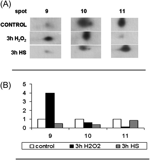
Fig. 5.
2-DE changes in the level of ascorbate peroxidase (APX) protein in cells exposed to heat shock and H2O2 treatments. (A) Immunoblotting analysis was performed using a specific APX antibody (see Materials and methods) in control cells, in cells treated at 55 °C for 10 min and recovered for 3 h (3 h HS), and in cells exposed to H2O2 for 3 h. (B) Densitometric analysis of antibody responses. The relative optical density is expressed in arbitrary units. The levels of three protein not differentially expressed (based on proteomic analysis) were used to normalize western blot signals. The image represents one of three independent replicas.

