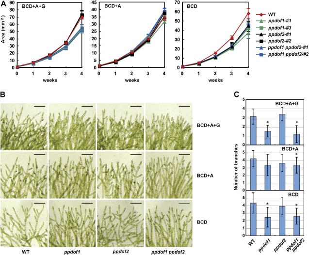
Fig. 3.
Nutrient condition-dependent phenotype of the disruptant lines. (A) Areas of protonemal colonies that were initially grown on regeneration medium for 4 weeks and then on BCD medium supplemented with ammonium and glucose (BCD+A+G), BCD medium supplemented with ammonium (BCD+A), or BCD medium for the indicated periods after protoplast formation. Values are the means ±SD of three replicates. (B) Images showing the branching of protonemal filaments of wild-type (WT), ppdof1 or ppdof2 single disruptant lines, and the ppdof1 ppdof2 double disruptant line. Scale bars=200 μm. (C) The numbers of branches within six cells from the filament top. These numbers were measured using protonemal colonies that were initially grown on regeneration medium plates for 4 weeks and then on BCD medium supplemented with ammonium and glucose (BCD+A+G), BCD medium supplemented with ammonium (BCD+A), or BCD medium for 3 weeks. Values are the means ±SD of 15 replicates. Asterisks indicate a statistically significant difference (P < 0.05), compared with the values obtained for wild-type P. patens.

