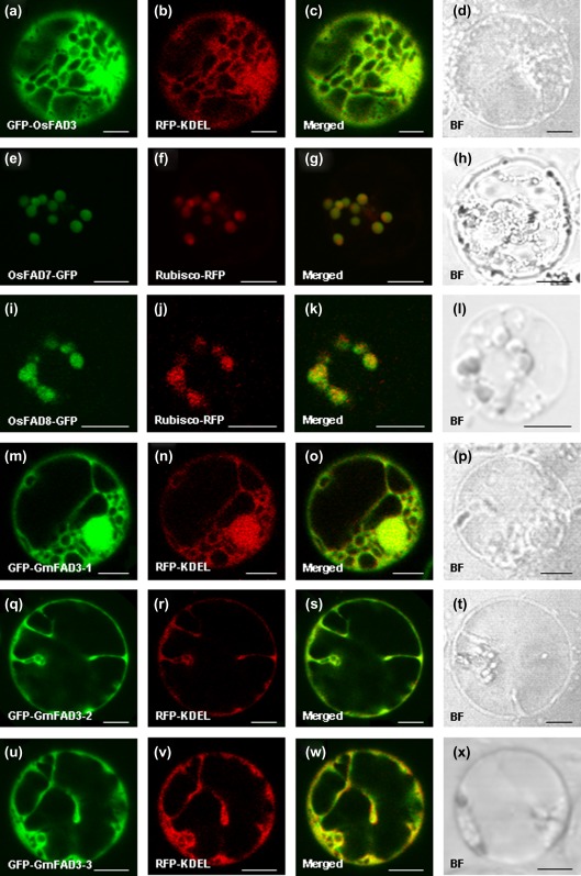
Fig. 1.
Subcellular localization of the OsFAD3/7/8 and GmFAD3-1/2/3 proteins. The green GFP and red RFP signals obtained by confocal microscopy indicate fusion proteins OsFAD7/8–GFP (e and i) or GFP–FAD3 (a, m, q, and u) and Rubisco–RFP (chloroplast marker protein; f and j) or RFP–KDEL (endoplasmic reticulum marker protein; b, n, r, and v) which were transiently expressed in rice protoplasts. The overlap of green and red fluorescent signals is indicated in merged images (c, g, k, o, s, and w). Bright fields are showed in d, h, l, p, t, and x. Bars, 5 μm.

