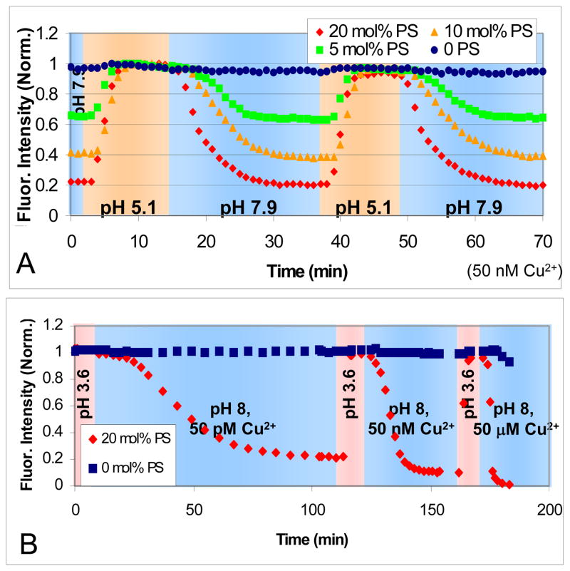
Figure 6.
In (A) the fluorescence intensity of 0, 5, 10, and 20 mole percent PS is shown as the pH was alternated between pH 5.1 (orange background) and 7.9 (blue background) in 1 mM citrate/MES/Tris with 50 nM Cu2+. (B) Experiments in which different concentrations of CuSO4 solution were tested at pH 3.6 (pink background) and 8.0 (blue background) with 50 pM, 50 nM, and 50 μM levels of CuSO4. The SLBs consisted of 0.1 mol% TR-DHPE in POPC at the labeled DOPS concentration. The buffer was 1 mM citrate/MES/Tris.

