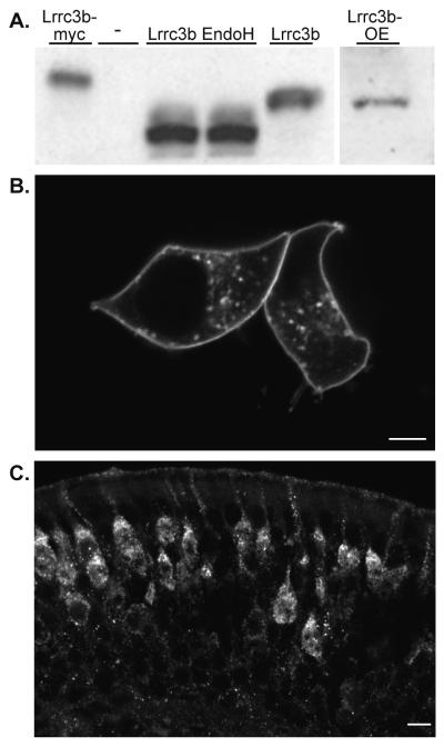
Figure 3.
Lrrc3b protein is localized to the plasma membrane and glycosylated (A) Purified anti-peptide Lrrc3b antisera (amino acids 47-64) recognizes a similar size (35kDa) protein in Lrrc3b-transfected HEK293 cell lysates (lane 5) and in native OE membrane preparations (lane 6). The antisera-reactive band migrates at a higher apparent molecular weight in lysates from myc-tagged Lrrc3b transfected cells (lane 1). Endoglycosidase H treatment of the Lrrc3b-transfected lysates (lanes 3 and 4) reduced the apparent size of the Lrrc3b product (25kDa) indicating the presence of glycosylation on the HEK-293 expressed and native protein. (B) HEK293 cells transfected with a Lrrc3b-YFP fusion protein, visualized by intrinsic YFP fluorescence, display intense signal at the plasma membrane and in a punctate pattern in the cytoplasm. scale bar = 5 microns (C) The native Lrrc3b protein, visualized with the peptide antisera is predominantly localized to dendrites and the apical peri-nuclear endoplasmic reticulum and Golgi regions. scale bar = 10 microns.

