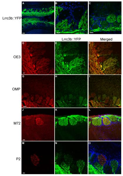
Figure 6.
YFP expression in the olfactory bulb of Lrrc3b(ΔYFP/+) mice. (A) YFP expression in the Lrrc3b(ΔYFP/+) OB. YFP is abundant in the olfactory nerve layer. Individual glomeruli display a wide range of signal intensity. DAPI staining of cell nuclei highlight the periglomerular cells that outline each glomerulus. The YFP signal is pseudocolored green. scale bar= 100 microns (B-C) High magnification images of olfactory glomeruli. Several strongly YFP-positive glomeruli are immediately adjacent to glomeruli that display no detectable YFP fluorescence. Regions with intermediate intensity signals are also observed. scale bar= 100 microns (D-I) All glomeruli receive innervation by ORNs based on the uniform presence of OE3 (D) or OMP (G). The variable intensity of Lrrc3b is apparent in intrinsic YFP fluorescence images of the Lrrc3b-tagged axons (E,H) and in the merged images (F,I). The OMP and OE3 signals were visualized using antibodies to lacZ in OMP(tau-lacZ/+) mice and spectral separation of GFP and YFP signals in OE3(tau-GFP/+) mice respectively. (J-O) Determination of the YFP florescence intensity of specific olfactory bulb glomeruli. Individual glomeruli innervated by M72- or P2-positive axons were identified with anti-β-gal antibody in M72(tau-lacZ/+) (J) and P2((tau-lacZ/+) (M) mice double heterozygous for the Lrrc3bΔYFP allele. The intense intrinisic YFP florescence of the glomerulus corresponding to M72 (K, L) suggests that cells expressing this OR are highly active. The glomerulus receiving input from P2-expressing ORNs are relatively inactive in the same odor environment (N,O). scale bars (D-O) = 100 microns

