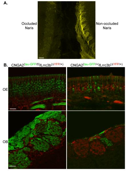
Figure 7.
Intrinsic fluorescence levels in the Lrrc3b(ΔYFP/+) mouse are altered by odor-evoked activity. (A) Fluorescence in a PD40 Lrrc3b(ΔYFP/+) mouse showing intrinsic signal in the two OBs examined near the midline. The signal is very low in the left OB of an animal subjected to a unilateral naris occlusion at PD2. The bulb from the non-occluded side (right side) displays robust YFP signal. scale bar= 100 microns (B) The OE of CNGA2(tau-GFP/+)/Lrrc3b(ΔYFP/+) female mice (upper left panel) contains an X-inactivation-induced mosaic of ORNs expressing exclusively GFP or YFP (pseudo-colored red in each panel). In the OB of these same mice (lower left panel), glomeruli are either GFP or YFP positive. Note the small size of the GFP-labeled CNGA2-deficient glomerulus. In the OE from male CNGA2(tau-GFP/0)/Lrrc3b(ΔYFP/+) mice (upper right panel), the ORNs are all inactive and uniformly express GFP but little or no YFP. The glomeruli in the OB (lower right panel) are uniformly GFP positive. YFP is undetectable in the glomerular neuropil but is present in periglomerular cells. scale bar (upper panels) = 25 microns. scale bar (lower panels) = 50 microns

