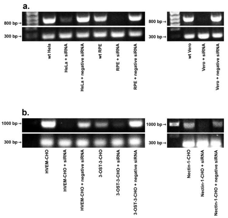
Fig. 3.

RT-PCR to verify reduced 2-OST gene expression. RT-PCR analysis of 2-OST expression was performed with HeLa, RPE, and Vero cells (a), and CHO-K1 cells expressing 3-OST-3B, HVEM, or nectin-1 (b). Cells were mock treated (wt) or transfected with scrambled siRNA (+ negative siRNA) or 2-OST siRNA (+ siRNA). About 48 h after siRNA transfection, total RNA was isolated from each cell line. Superscript II reverse transcriptase was used for RT-PCR. PCR amplification of cDNA was done using specific 2-OST primers. Expected PCR product sizes were 792 bps (2-OST) and 285 bps (β-actin) (bottom panels).
