
Fig. 5.
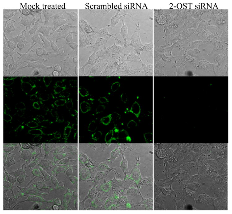
Use of immunofluorescence to confirm reduced 3-OS HS expression. HeLa cells were incubated with the antibody HS4C3 for 1 h at 4 °C. Cells were then fixed for 20 min, and incubated with FITC conjugated anti-mouse IgG to label 3-OS HS surface expression. 3-OS HS surface expression was compared in HeLa cells that were mock treated (left panels) or transfected with scrambled siRNA (middle panels) or 2-OST siRNA (right panels). Imaging was performed using confocal microscopy at a 60x oil objective.