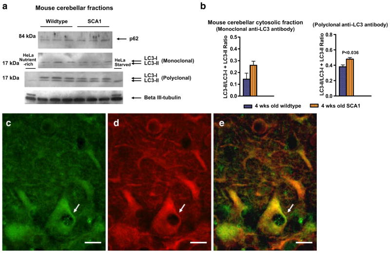
Fig. 6.
a Western blots of p62, LC3, and βIII-tubulin in mouse cerebellar fractions of 4-week-old wild-type (n=3) and SCA1 (n=3) mice. Samples were subjected to SDS-PAGE, followed by electro-transfer and Western blot analysis. Western blots analysis of LC3 using mono- and polyclonal antibodies, which recognize both LC3-I and LC3-II forms (Medical & Biological Laboratories, MBL International Corp.) showed altered levels of LC3-I and LC3-II in SCA1 mice. HeLa cell lysates prepared from cells either incubated with 10% fetal calf serum or starved overnight were used as internal controls to identify LC3-II bands (Medical & Biological Laboratories). The data presented in b are the mean±SD of LC3-II/(LC3-I + LC3-II) ratios calculated from the band intensity measurements of the Western blots. Statistical significance was calculated using GraphPad Prism, unpaired Student’s t test for independent samples, version 4.0 (GraphPad Software). A value of P<0.05 was accepted as statistically significant. c–e Double immunofluorescence showing LC3 (green, c) and βIII-tubulin (red, d) immunostaining in PCs in a 4-week-old SCA1 mouse. e Merged image. Arrows indicate PC vacuoles containing LC3. Bars=10 μm

