Fig. 8.
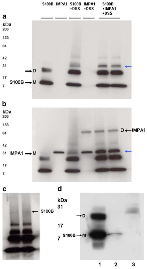
a–c Western blots showing cross-linking of S100B with IMPA1. Purified bovine brain S100B protein and IMPA1 were incubated in PBS in the presence and absence 0.5-mM protein cross-linker DSS, and then subjected to SDS-PAGE followed by Western blot analysis. The same amount of proteins was used in all experiments and loaded in all lanes. a Blot probed for S100B. This protein under reducing conditions exists as a monomer (M) of 11 kDa, a dimer (D), and as a multimer. b Blot reprobed for IMPA1. IMPA1 normally exists as a dimer, but under reducing conditions appear as a monomer with approximate molecular weight of 30 kDa (black arrow); c overexposed blot a showing higher molecular weight S100B band corresponding to IMPA1 dimer in b. Blue arrows in a and b indicate the position of S100B. d Western blot of S100B showing co-immunoprecipitated S100B with IMPA1 (lane 2). IMPA1 antibody bound to protein A agarose beads was used to co-immunoprecipitate S100B from IMPA1–S100B protein mixture as described in the “Materials and Methods” section. Lane 1 shows standard S100B protein. Lane 3 Instead of IMPA1 antibody, control serum was used

