Figure 3.
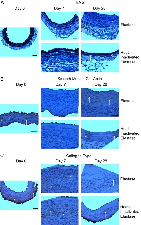
Histology and immunohistochemistry. Histological and immunohistochemical characterization of elastin (A), smooth muscle cell actin (SMCA) (B), and adventitial collagen type I (C) validated immunofluorescent array tomography (IAT) findings. (A) Elastic van Gieson (EVG) results depict the loss of elastin thickness and organization and the increase in non-adventitial wall thickness that occurred during aneurysm development in the elastase group. Elastin lamellae structure and organization in the heat-inactivated elastase group were relatively constant. Arrows highlight elastin stained by EVG. (B) SMCA content changed dynamically with a decrease from day 0 to day 7 and subsequent increase from day 7 to day 28 in both the elastase and heat-inactivated elastase groups. Arrows highlight regions with positive staining for SMCA. (C) In both the elastase and heat-inactivated elastase groups, adventitial collagen type I content at days 7 and 28 decreased relative to day 0. Arrows highlight areas with positive staining for collagen type I. Orientation of all images: lumen at top and adventitia at bottom of image. Images taken at 63× magnification with scale bar representing 25 µm.

