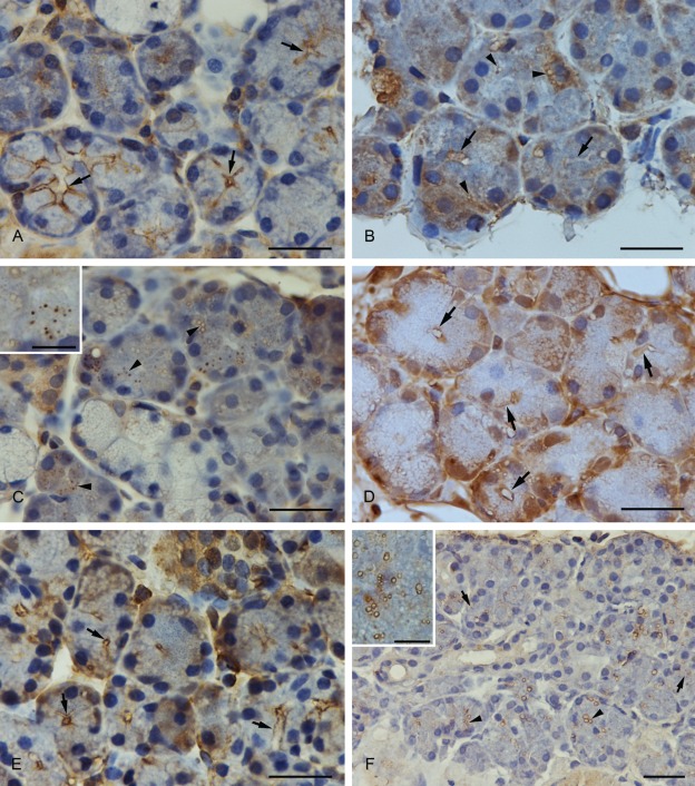
Figure 2.
(A) Anti-SNAP-23 immunostaining. SNAP-23 was concentrated at the luminal and the basolateral membrane (arrows). (B) Anti-syntaxin-2 immunostaining. Syntaxin-2 showed a strong expression at the apical plasma membrane (arrows) and in smaller and larger vesicles (arrowheads). The basal part of the cell, especially the perinuclear compartment, was also strongly stained. (C) Anti-syntaxin-2 immunostaining. Larger vesicles with an unstained matrix and smaller completely stained vesicles showed a positive syntaxin-2 immunostaining. A higher magnification of the vesicles is demonstrated in the inset. (D) Anti-syntaxin-4 immunostaining. Syntaxin-4 was expressed at the apical and basolateral plasma membrane of submandibular acinar cells (arrows). (E) Anti-VAMP-2 immunostaining. VAMP-2 was localized at the apical region of the plasma membrane (arrows). (F) Anti-septin-2 immunostaining. Positively stained vesicles with an unstained matrix (arrowheads) and completely stained vesicles (arrows) were visible with the antibody to septin-2. A higher magnification of the vesicles is demonstrated in the inset. Scale bars: 25 µm; scale bars in insets: 6 µm.

