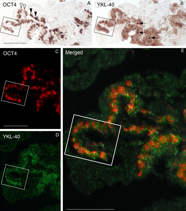
Figure 2.
Staining patterns for OCT4 (A), YKL-40 (B), and OCT4/YKL-40 (C–E) in three consecutive sections of a human embryonic stem cell colony (LRB03) from culture day 0 visualized by immunohistochemistry and confocal fluorescence microscopy. The “U-turn” in the boxed area is easy to identify in the three sections. (A) All cell nuclei exhibit OCT4 positivity, although a varying intensity of the staining is evident. Open arrowheads point to weakly stained nuclei, and solid arrowheads show strongly labeled nuclei. (B) The adjacent section stained for YKL-40 shows a marked reactivity in the cytoplasm with occasionally denser areas (arrows). (C–E) Confocal microscopy of a neighboring section stained simultaneously with antibodies against OCT4 and YKL-40. Cells with nuclear reactivity (red) for OCT4 (C) also exhibit cytoplasmic reactivity (green) for YKL-40 (D), verified at higher magnification by the merged image (E). Yellow dots correspond to YKL-40 located directly below or above the OCT4-positive red nucleus. Scale bars: 100 µm.

