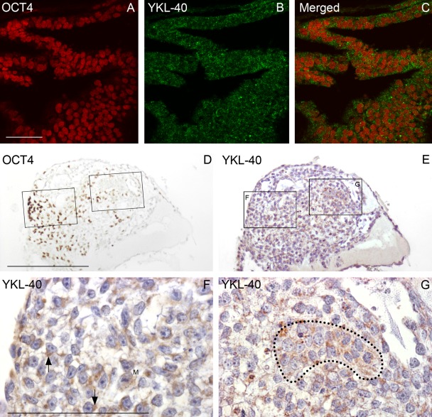
Figure 3.
Early and later differentiating colonies. (A–C) Confocal microscopy of a 7-day-old colony from LRB03 grown without basic fibroblast growth factor shows early differentiating human embryonic stem cells (hESCs) stained for OCT4 (A) and YKL-40 (B). The merged picture (C) depicts strongly OCT4-reacting stem cell nuclei surrounded by a fine granular cytoplasmic YKL-40 staining. A later differentiating colony (shown in D–G) in bright-field microscopy is from a 17-day-old culture from LRB010 also stained for OCT4 (D) and YKL-40 (E). In (D), a small group of strongly OCT4-positive cells is present toward one edge of the colony with few weakly OCT4-positive cells scattered toward the middle. A neighboring section stained for YKL-40 (E) with boxed areas shown at higher magnification in (F) and (G) depicts YKL-40 in the cytoplasm of loosely packed cells, often in close proximity to the nucleus (arrows in F). Morphology of the individual cells is corresponding well to hESC morphology with large nuclei with one to three dense nucleoli in a scanty cytoplasm. However, the cell pattern is less epithelial and more mesenchymal. In the rather OCT4-negative field in (G), the epithelial-like cell group marked by a dotted line shows a dense granular cytoplasmic YKL-40 reactivity and nuclei without dense nucleoli. M shows a mitotic figure. A–C: same magnification, scale bar: 50 µm. D, E: same magnification, scale bar: 250 µm. F, G: same magnification, scale bar: 100 µm.

