Figure 4.
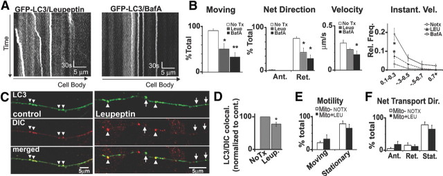
Lysosomal proteolysis inhibition slows LC3 vesicle transport without causing generalized axonal transport defects. A, Representative 5 min kymographs of GFP-LC3 movies after leupeptin (20 μm, 24 h) or bafilomycin A (10 nm, 2 h; compared with controls) (see Fig. 2A). B, Quantification of GFP-LC3 movements after leupeptin (n = 81 vesicles), bafilomycin A (n = 44 vesicles), or controls (n = 96 vesicles). The percentage of moving LC3 vesicles (motility) and LC3 vesicles undergoing net retrograde movement are significantly reduced by leupeptin or bafilomycin treatment. LC3 vesicles also have slower retrograde velocities after leupeptin or bafilomycin, and the frequency of instantaneous retrograde velocities show a depression after leupeptin or bafilomycin. C, DIC immunolabeling on GFP-LC3 vesicles under normal conditions and after treatment with leupeptin. A portion of LC3 vesicles did not colocalize with DIC after leupeptin (arrowheads). D, Quantification of the percentage of LC3 vesicles that colocalized with DIC (per axon) shows reduction after leupeptin (n = 60). E, F, Motility (E) and net direction (F) of DsRed-Mito-positive mitochondria (control, n = 69; leupeptin, n = 78) show that mitochondria movements were not affected by leupeptin (20 μm, 24 h). Values represents means ± SEM. *p < 0.05; **p < 0.01. No Tx, No treatment; BafA, bafilomycin A; Leup or LEU, leupeptin.

