Figure 6.
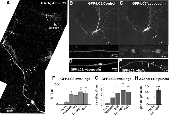
Lysosomal proteolysis inhibition accumulates LC3 vesicles. A, Endogenous LC3-immunoreactive vesicles (arrows) are abundant and accumulated (arrowhead) after treatment with bafilomycin (10 nm, 4 h). The cell body contains relatively few LC3 puncta compared with the axon. B, C, Representative live image of GFP-LC3 neuron before (B) or after (C) treatment with leupeptin (20 μm, 24 h). LC3 vesicles accumulate in swellings (arrows). The enlarged area of axonal swellings is shown below. D, E, Live image of axonal GFP-LC3 vesicles after treatment with leupeptin (20–40 μm, 24 h; D) or bafilomycin A (50 nm, 4 h; E) where vesicles are both dispersed (arrows) and accumulated in a focal swelling (arrowhead). F, Quantification of the percentage of GFP-LC3 neurons with neuritic swellings containing GFP-LC3. G, The number of GFP-LC3 vesicle-enriched swellings per millimeter length of neurite after treatment with various protease inhibitors (see Materials and Methods). H, Quantification of the number of GFP-LC3 vesicles per 100 μm in control or after leupeptin treatment (n = 50). The frequency of GFP-LC3 vesicles increases ∼2.5-fold after leupeptin. Values represents means ± SEM. All scale bars are as indicated. **p < 0.01; ***p < 0.0001. No Tx, No treatment; BafA, bafilomycin A.

