Figure 7.

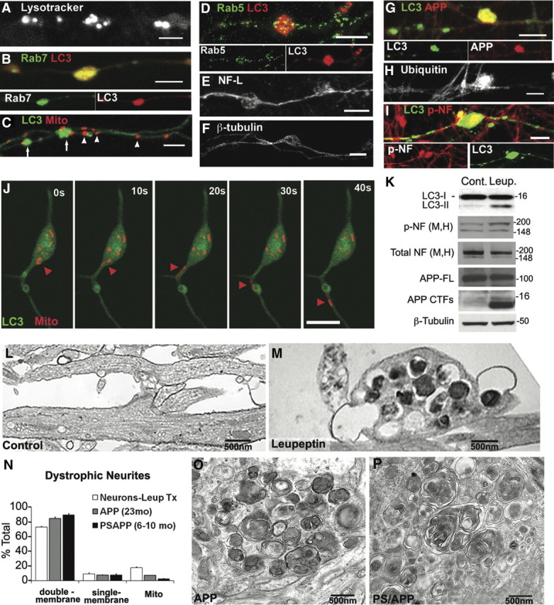
Biochemical and ultrastructural profiles of neurites after lysosomal clearance inhibition resemble AD dystrophic neurites. A–F, Swellings preferentially accumulate with lysosomal (proteolytic) vesicles after treatment with leupeptin (20 μm, 24 h). LysoTracker Red vesicles (A), Rab7 vesicles (B), and LC3 vesicles (C, arrows) are preferentially accumulated, whereas mitochondria (C, arrowheads), Rab5-positive early endosomes (D), neurofilament-light chain (E), or β-tubulin (F) are relatively evenly distributed along the axon. G–I, Swellings accumulate proteins that identify AD-dystrophic neurites including APP-containing autophagic vesicles (G; C1/6.1 antibody for C terminus of APP double labeled with LC3), ubiquitinated proteins (H), and phospho-neurofilaments (I; NF-M/H; double labeled with LC3). Phosphorylated neurofilament accumulation occurs in swollen regions containing accumulated LC3 vesicles. Scale bars, 5 μm. J, Time lapse of swelling with GFP-LC3 and DsRed-Mito cotransfection and treatment with leupeptin (20 μm, 5 h). The cell body is at the top. Although both GFP-LC3 vesicles and DsRed-mitochondria are accumulated, mitochondria occasionally resume transport (red arrowheads), whereas LC3 movement out from the swelling is not observed. K, Western blots with indicated antibodies in leupeptin-treated neurons compared with controls. Leupeptin increases the ratio of LC3-II/I, phospho-neurofilaments (SMI-31), and APP-CTFs, without increasing overall levels of neurofilaments or APP holoprotein (full length). Molecular mass is shown in kilodaltons. L, M, Representative electron micrographs of neurites after leupeptin (20 μm, 24 h; M) compared with controls (L). Leupeptin-induced accumulation of double-membrane, amorphous electron-dense AVs in neurites is shown. N, Quantification of morphometric analysis of organelles in dystrophic neurites from leupeptin-treated neurons (Leup Tx; n = 20) or AD mouse models (APP, n = 25; PSAPP, n = 25). O, P, Representative electron micrographs of dystrophic neurites from APP (O) and PSAPP (P) mouse brain. Organelles within dystrophic neurites are mostly double-membrane AVs. Values represents means ± SEM. Scale bars: A–J, 5 μm.
