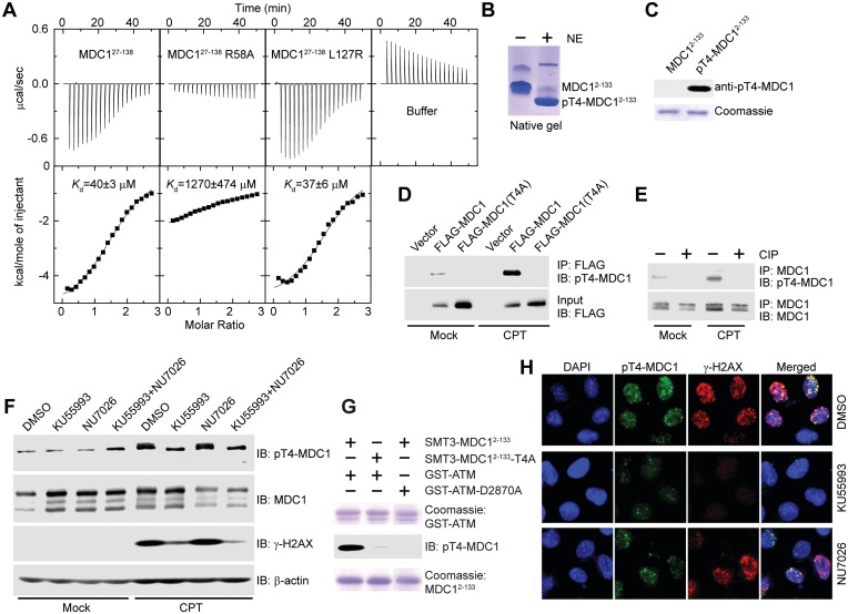
Figure 3.
MDC1 T4 is phosphorylated by ATM in response to DNA damage, and pT4 is recognized by the FHA domain. (A) Microcalorimetric titration of phosphopeptide pT4-8P into MDC127–138, its mutants R58A and L127R, or 20 mM sodium phosphate (pH 7.6) and 150 mM NaCl buffer. The heat peaks were integrated, corrected for the ligand dilution effect and fit to a one-set-of-sites binding model. The dissociation constant, Kd, values are indicated. (B) MDC12–133 was phosphorylated by nuclear extracts (NE) of HeLa cells and resolved in a native gel. (C) Affinity purified rabbit polyclonal anti-pT4-MDC1 antibody specifically recognizes phosphorylated MDC12–133 but not unphosphorylated MDC12–133. (D) pT4 in transiently expressed MDC1. Empty vector or vectors expressing FLAG-MDC1 or FLAG-MDC1(T4A) were transfected into U2OS cells. The cells were mock treated or treated with CPT for 1 h, followed by immunoprecipitation with anti-FLAG antibody and immunoblotting with anti-FLAG and anti-pT4-MDC1 antibodies. (E) pT4 in endogenous MDC1. U2OS cells were mock treated or treated with CPT for 1 h. The total cell lysates were immunoprecipitated with anti-MDC1 antibody, treated with or without calf intestine phosphatase (CIP), and blotted with anti-pT4-MDC1 and anti-MDC1 antibodies. (F) T4 is primarily phosphorylated by ATM. U2OS cells were treated with DMSO, ATM inhibitor KU55993 (10 μM), DNA-PKcs inhibitor NU7026 (10 μM) or both KU55993 and NU7026 for 2 h before treatment of 0 or 10 μM CPT for 1 h. The total cell lysates were resolved in SDS-PAGE and blotted with anti-pT4-MDC1, anti-MDC1, anti-γ-H2AX and anti-β-actin antibodies. Endogenous MDC1 presents multiple isoforms when blotted with anti-MDC1 antibody; only the slowest migrating one was recognized by anti-pT4-MDC1 antibody. (G) SMT3-MDC12–133, but not its T4A mutant, can be phosphorylated in vitro by a recombinant GST-fused kinase domain of ATM (GST-ATM, residues 2709–2964). GST-ATM-D2870A is a kinase-dead mutant. (H) U2OS cells were treated as in (F) and were immunostained with anti-pT4-MDC1 (green) and γ-H2AX (red). DNA was stained by DAPI (blue).

