Figure 7.
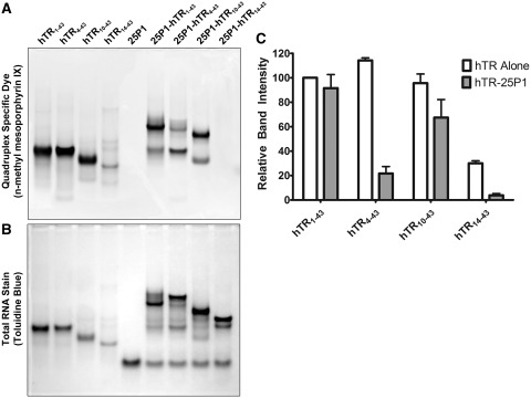
A switch between an internal and terminal quadruplex occurs upon P1 helix formation. (A) hTR1–43 and successive 5′ truncations were heated to 95°C either alone or in the presence of a 2-fold molar excess of 25P1 for 5 min and then allowed to cool to room temperature. Approximately 500 pmols of each RNA was separated by native TBE polyacrylamide gel electrophoresis and stained with the quadruplex-specific fluorescent dye n-methyl mesoporphyrin IX. A significant decrease in staining intensity was observed for hTR14–43 indicating a loss of quadruplex structure. The hTR1–43-25P1 complex stained with the quadruplex-specific dye; however, this staining was completely lost in the hTR14–43-25P1 complex. (B) Following fluorescent visualization of the gel it was further stained with the total RNA stain toluidine blue. (C) Densitometry quantification of the bands in (A). In the case of hTR-25P1 complexes, the dominant band, as observed in (B), was chosen for quantification. Data represents the mean of three independent experiments ± standard deviation. Additional gel images are provided in Supplementary Figure 7.

