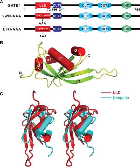
Figure 1.
Structure of the SATB1 ULD. (A) Schematic representation of the domain organization of mouse SATB1. The ULD domain boundary identified in this work is located from Gly71 to Ser172, and a novel CUTL domain is located from His186 to Lys244. The two mutants used in this study, KWN–AAA and EFH–AAA, were created by substituting the 136K137W138N and the 97E98F162H motifs with ‘AAA’ cassettes. (B) Cartoon representation of the overall structure of ULD. The N- and C-termini of the protein are labeled. (C) Stereo view showing the superimposed structures of ULD and ubiquitin (PDB code: 1UBI).

