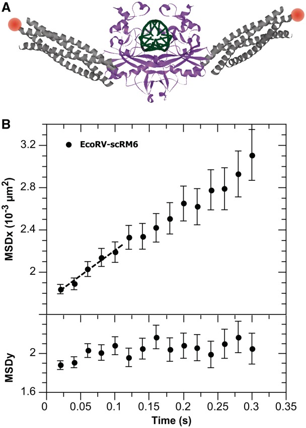
Figure 2.
(A) One possible model of EcoRV (protein data bank 4rve) fused to scRM6 protein (protein data bank 1qx8), in which the structure of scRM6 protein is aligned with the N-terminal helix of EcoRV. EcoRV is presented in magenta, DNA in green, scRM6 protein in gray and the label in orange. (B) The longitudinal (along the DNA molecule) and transverse (perpendicular to the DNA molecule) MSD of EcoRV fused to the scRM6 protein labeled with Cy3B. The longitudinal MSD depends linearly on time, which shows that the fusion protein slides along the DNA, while, as expected, the transverse MSD is constant over time. The linear diffusion constant D is derived from the slope of the curve (dashed line: linear fit on the first five points of the MSD) using the relation: slope = 2D.

