
FIGURE 9.
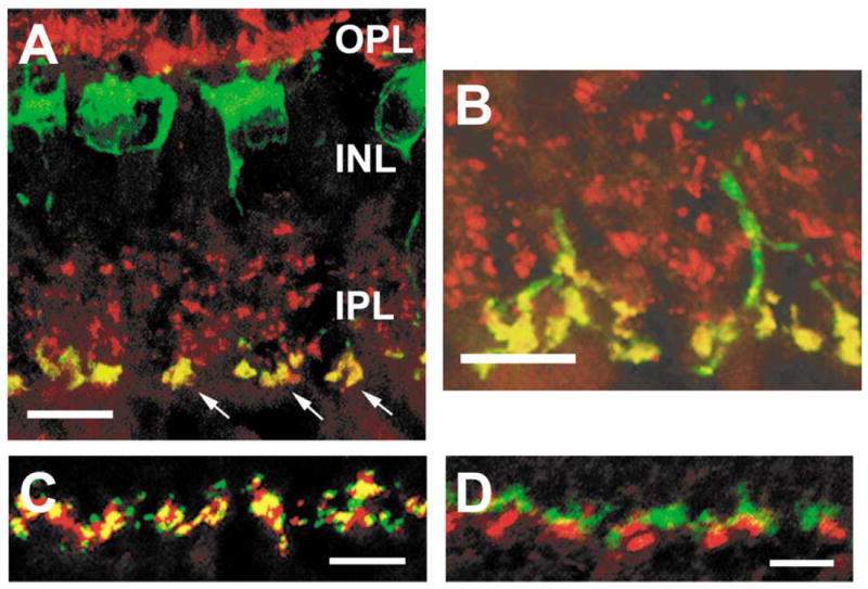
Serotonin receptors in the rabbit retina. (A-B) Serotonin receptors of the 5-HT2A subtype are localized to rod bipolar cell terminals in the rabbit retina. These vertical sections were labeled with antibodies to 5-HT2A (red) and protein kinase C (PKC, green). 5-HT2A-IR puncta were found in the inner plexiform layer (IPL, arrows) (A) and outer plexiform layer (OPL) (B). All of the PKC-IR rod bipolar cells expressed 5-HT2A in both the IPL and OPL. (C-D) 5-HT3A receptors in the rabbit retina are localized to rod spherules in the outer plexiform layer (OPL). Using an antibody to 5-HT3A (red), large puncta were labeled in the OPL. The yellow regions in panel (C) demonstrate that the 5-HT3A-IR were colocalized with a marker for rod spherules, B16 (green). Alternatively in panel (D), a marker of cone pedicles, peanut agglutin (green), was not associated with 5-HT3A-IR puncta. (A-B) Modified and reprinted from Pootanakit and Brunken.122 (C-D) Modified and reprinted from Pootanakit et al.117