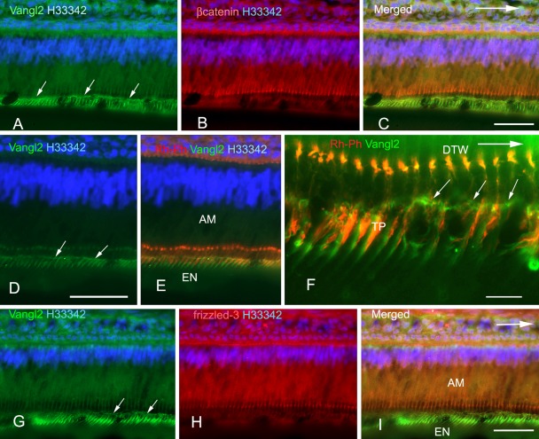
Figure 2.
Triple labeling of inner enamel–secretory ameloblasts with anti-Vang12 and Hoechst 33342 (H33342) and anti–β-catenin, rhodamine-phalloidin, or anti–frizzled-3. Sections of rat incisors were stained with anti-Vang12 Ab (A, C–G, I) (green), anti–β-catenin (B, C) (red), rhodamine-phalloidin (Rh-Ph) (E, F) (red), or anti–frizzled-3 (H, I) (red), and nuclei were stained with Hoechst 33342 (H33342) (A–E, G–I) (blue). A merged image of (A) and (B) is shown in (C). An anti-Vang12 Ab-labeled horizontal fluorescent line (arrows in A, D, F, G) was localized at the proximal part of Tomes’ processes. This region was located 5 to 10 µm below the distal junctional complexes that were labeled with anti–β-catenin (B, C) or F-actin (E, F). Anti–frizzled-3 antibodies labeled distal junctional complexes (H, I). Anti-Vang12-labeled vertical fluorescent lines were also seen below the horizontal fluorescent line in Tomes’ processes (A, C–G, I). Double-labeling of Tomes’ processes (TP in F) with anti-Vang12 Ab and rhodamine-phalloidin showed that these vertical fluorescent lines were localized at the lateral surface of Tomes’ process on the apical side of the incisor (F). The arrows indicate the incisal direction. AM, ameloblast; EN, enamel; DTW, distal terminal web; TP, Tomes’ process. (C, D, I) Bars = 50 µm. (F) Bar = 10 µm.

