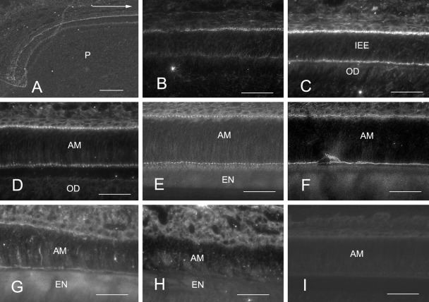
Figure 4.
Frizzled-3 labeling of freeze-dried sections of rat incisors without fixation and demineralization. Anti–frizzled-3 labeling (A–H) of inner enamel epithelia and ameloblasts at the following stages are shown: proliferation (A), early differentiation (B), late differentiation (C), early inner enamel secretion (D), middle inner enamel secretion (E), outer enamel secretion (F), transition (G), and early maturation (H). A section for negative control is also shown (I). Initially, the proximal part of the inner enamel epithelia was anti–frizzled-3 Ab-positive (A, B). The distal part then became positive in late differentiation (C). Both proximal and distal junctional complexes in the region of inner and outer enamel secretion were positive (D–F), but fluorescence gradually diminished at the transition zone (G) and disappeared in the zone of maturation (H). The section for negative control showed no specific labeling (I). Odontoblasts showed no labeling with the anti–frizzled-3 Ab (C, D). The arrow in (A) shows incisal direction. IEE, inner enamel epithelial cell; AM, ameloblast; EN, enamel; OD, odontoblast; P, dental pulp. Bars = 50 µm.

