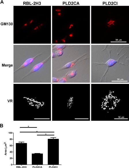
Figure 3.
PLD2CI cells have alterations in the Golgi complex. In RBL-2H3 and PLD2CA cells, the Golgi complex was compact and localized juxtanuclearly, but in PLD2CI cells, the Golgi complex was more diffuse and spread throughout the cytoplasm. Volume rendering (VR) of the Golgi complex confirmed the differences in the size of the Golgi complex among the three cell lines. (B) Quantification of the area of the Golgi complex showed that PLD2CA cells had a smaller Golgi complex than the RBL-2H3 cells (*=p<0.05). The PLD2CI cells had a larger Golgi area when compared with the RBL-2H3 cells (*=p<0.05). GM130, red; DAPI, blue.

