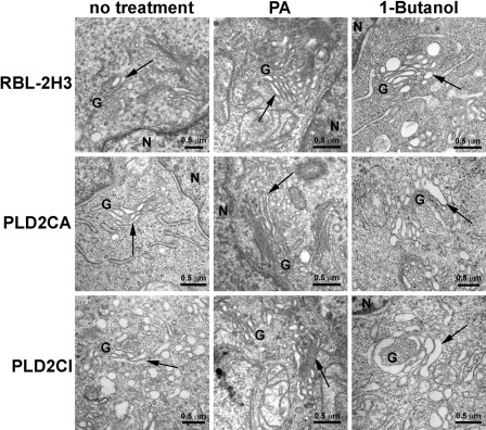
Figure 4.
By transmission electron microscopy (TEM), the RBL-2H3 and PLD2CA cells had a well-organized Golgi complex, with their cisternae showing the typical arrangement of flattened saccules (arrows). The Golgi complex of the PLD2CI cells was dispersed in the cytoplasm, and the cisternae were disorganized and dilated (arrow). There was also an increase in the number of Golgi-associated vesicles in the PLD2CI cells. By TEM, treatment of RBL-2H3 cells with 10 mg/ml PA for 24 hr did not alter the morphology of Golgi complex. However, treatment with 1% 1-Butanol for 20 min led to the disorganization of the Golgi complex and dilated cisternae. Treatment of PLD2CA cells with phosphatidic acid (PA) did not alter the organization of the Golgi complex. In these cells, treatment with1% 1-Butanol also resulted in the disorganization of the Golgi complex and dilation of the cisternae. Treatment with PA in PLD2CI cells led to a decrease in the size of the Golgi complex cisternae and number of Golgi-associated vesicles (arrows), but treatment with 1% 1-Butanol did not alter the structure of the Golgi complex (arrows). G, Golgi complex; N, nucleus.

