Figure 1.
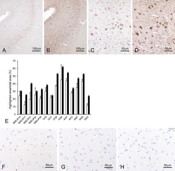
Comparison of immunolabeling intensity by the conventional polymer method and the tyramide signal amplification (TSA) biotin system. Localization of the disease-associated prion protein (PrPSc) in the dorsolateral prefrontal cortex along the superior frontal sulcus of bovine spongiform encephalopathy (BSE)–affected cattle (case 4437) by using mAb F99/97.6.1 (A–D). The graph illustrates the mean percentage of the ratio of PrPSc-immunolabeled area to total area (highlighted area/total area [%]) for each animal analyzed by using ImageJ (E). No statistically significant differences were determined between TSA (filled bars) and conventional polymer (empty bars) methods (Student’s t-test). Data expressed as the means ± standard deviation. Weak background immunostaining is present with the TSA system (F) but not the conventional polymer method (G) in the cerebral cortex of an uninfected control animal immunolabeled with mAb F99/97.6.1. No specific background immunolabeling is detected in the cerebral cortex of BSE cattle (case 4394) immunostained with phosphate-buffered saline instead of the primary prion protein (PrP)–primary antibody using the TSA system (H).

