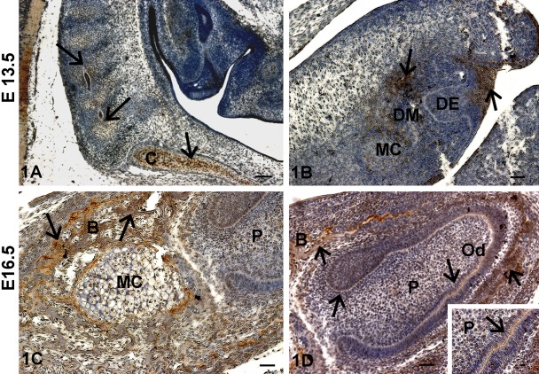
Figure 1.
Localization of TRIP-1 during embryonic development of bone and teeth. (A) Localization of TRIP-1 in the chondrocytes of the rostral part of the vertebral column with the forming cervical vertebrae. (B) Localization of TRIP-1 in the bud stage of tooth development. (C) Localization of TRIP-1 in Meckel’s cartilage (MC). (D) Localization of TRIP-1 in the developing incisors and alveolar bone at E16.5. Insert shows higher magnification of 1D showing TRIP-1 localization in the basement membrane. Black arrows in all images represent localization of TRIP-1. DM = dental mesenchyme; DE = dental epithelium; B = bone; P = dental pulp cells; C = chondrocytes in the cartilaginous primordium of the basioccipital bone; Od = odontoblasts. A–D bars = 50 µm; inset bar = 10 µm.

