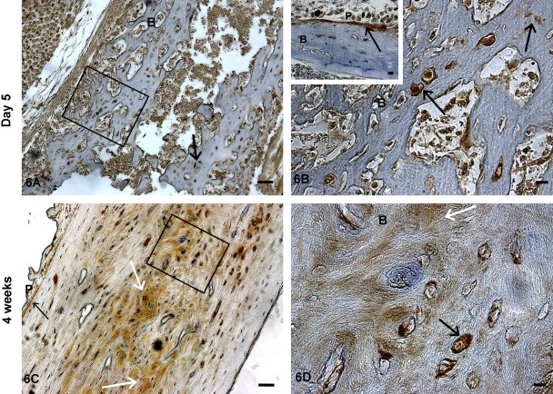
Figure 6.
Localization of TRIP-1 in postnatal day 5 and 4-week-old long bones of developing mice. Localization of TRIP-1 in the long bone sections of postnatal day 5 (A) and 4-week-old (C) mice. (B, D) Enlarged images show the presence of TRIP-1 in the mineralizing matrix and in osteocytes, osteoblasts, and periosteum. Insert in Fig. 6B shows the expression of TRIP-1 in the peroisteum. Black arrows point to expression of TRIP-1 in the bone osteocytes and periosteum. White arrows represent expression of TRIP-1 in the ECM. B = bone; P = periosteum. A,C bars = 50 µm; B,D, and inset bars = 10 µm.

