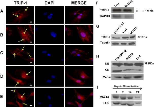
Figure 7.
Immunolocalization of TRIP-1 in different cell types. Figures 7A–E represent confocal images of the immunolocalization of TRIP-1 in different cell types: (A) primary odontoblasts; (B) primary osteoblasts; (C) human marrow stromal cells; (D) T4–4 preodontoblasts; and (E) MC3T3-E1 preosteoblast cells. Localization of TRIP-1 is represented by yellow arrows. Scale bars = 10 µm. (F) Northern blotting analysis of T4–4 preodontoblasts and MC3T3-E1 preosteoblast cells showing expression of TRIP-1. The size of TRIP-1 transcript obtained was 1.6 kb. (G) Western blot analysis of total protein lysates from C3H10T1/2 undifferentiated mesenchymal cells, T4–4 preodontoblasts, MC3T3-E1 preosteoblasts with TRIP-1 antibody showing expression of TRIP-1 at 37 KDa. (H) Western blot analysis of nuclear, cytoplasmic, and the secretome of the above 3 cell types shows distinct presence of TRIP-1 at 37 KDa. (I) Western blot analysis of proteins isolated from the secretory pool before (day 0) and after inducing differentiation (days 7, 14, and 21) for both T4–4 and MC3T3 cells showing the presence of TRIP-1. A-E TRIP-1, DAPI, and MERGE bars = 10 µm.

