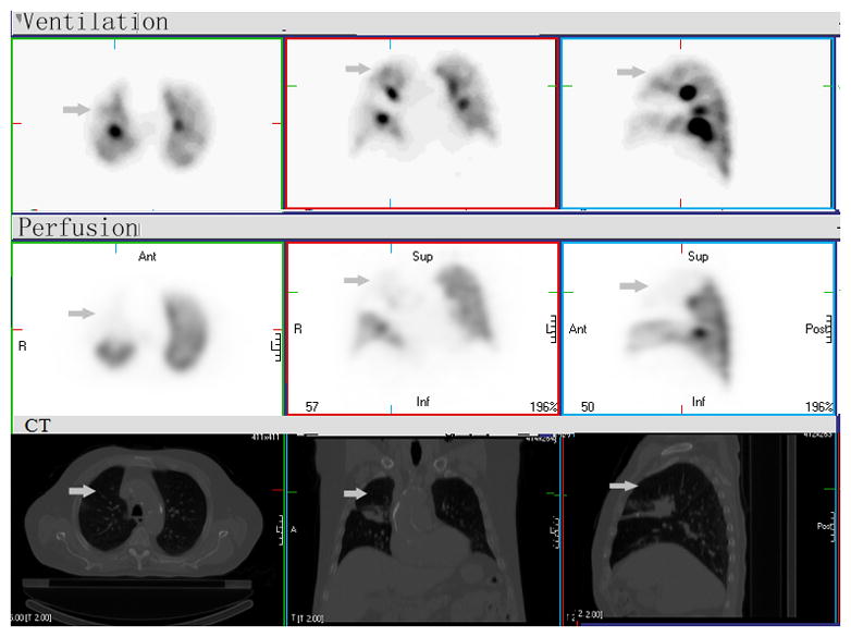
Figure 3. Mismatched V/Q defect.

There is a large mismatched defect with significantly reduced perfusion (Q) and normal V (arrow) in right middle and upper lobes, consistent with extrinsic compression of the pulmonary artery by the mass in hilum and mediastinum. If based on Q-SPECT only, this mismatched region would be considered as defect (“type B3” region) and potential for high dose radiation. Combined V/Q SPECT-CT can define and apply these regions as “type C region”, normal functioning “good” lung. The RT dose to such region should be minimized to decrease functional or clinically significant sequalae.
