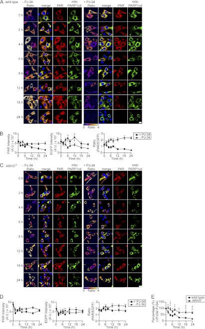
FIGURE 7.
Absence of ARH3 abolishes degradation of matrix-accumulated PAR. A and C, wild-type and ARH3−/− MEFs transiently expressing mitoPARP1cd were incubated in the absence (left panels) or presence (right panels) of 5 μm PJ-34 for the indicated times and then subjected to PAR immunocytochemistry. Upon PARP inhibition, the mitochondrial PAR content decreased over time in mitoPARP1cd-expressing wild-type MEFs but persisted in ARH3−/− MEFs. Scale bar = 20 μm. B and D, graphs summarize the time course of PAR signal and EGFP fluorescence intensities and PAR/EGFP signal ratio in the presence (●) and absence (▴) of 5 μm PJ-34 (n = 17–50 cells for each time point). Data are representative of three experiments. E, summarized data of relative PAR/EGFP signal ratios in wild-type (●) and ARH3−/− (▴) cells. Values were calculated by dividing the PAR/EGFP signal ratio in the presence of PJ-34 by that in its absence. Mean ± S.E. *, p < 0.05; **, p ≤ 0.01; ***, p ≥ 0.001 (two-way repeated measures analysis of variance with post hoc Bonferroni test).

