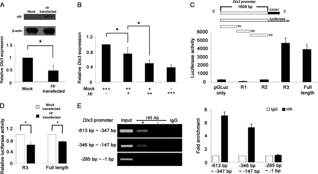
FIGURE 4.
HR down-regulates Dlx3 mRNA in Hr-transfected mouse keratinocyte. A, Western blot analysis showing the HR protein expressed in Hr-transfected PAM212 cells. β-Actin indicates equal amount of protein loading (top). Down-regulation of Dlx3 mRNA by HR in Hr-transfected PAM212 cells, as determined by real time PCR (bottom). B, Dlx3 was down-regulated by HR in a dose-dependent manner. +, 1 μg of DNA used for transfection. C, schematic representation of Dlx3 promoter construct for reporter assay. Promoter activities of the full length (1608 bp), R1, R2, and R3 clones of Dlx3 were compared with that of the pGLuc-vector. D, both R3 and full-length (1608 bp) Dlx3 promoter activities were decreased by HR expression. The Dlx3 promoter-fused reporter gene was transfected with the expression vectors of either Hr or pcDNA 3.1. Relative luciferase activity was normalized against transfection efficiency determined by β-galactosidase activity. Asterisks indicate p < 0.05. A–D, the activity was the average of three independent experiments conducted in duplicate (mean ± S.D.). E, ChIP analyses of HR on Dlx3 R3 promoters. HR binds the Dlx3 promoter in the region spanning −613 to −286 bp but not −285 to −1 bp. No antibody and normal IgG were used for the control experiment (left panel). Fold enrichment of HR against IgG was quantified using real time PCR performed in duplicate of three repeat experiments (right panel).

