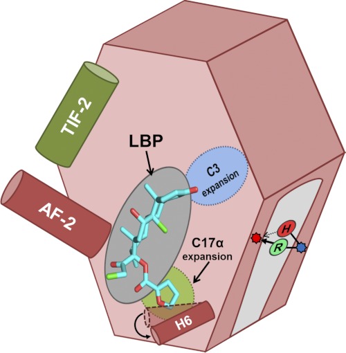
FIGURE 6.
Schematic summarizing relevant features of SR LBP that dictate MOF activation. Activation occurs when a ligand (e.g. MOF (cyan)) binds to the LBP, stabilizing the AF-2 helix (dark red) to allow for coactivator binding (e.g. TIF2 (dark green)) and subsequent transcriptional control. In GRs and PRs, the LBP can expand to accommodate steroids that are substituted at C3 (blue) or C17α (light green). In MR, we identified a single site outside of the LBP that can toggle MOF agonism versus antagonism, ostensibly by forming a bridge between H7 (blue dot) and the H5-β1 loop (red dot). In wild-type MR, His-853 makes a weak hydrogen bond that cannot support MOF activation (red H); the historical substitution to a positively charged arginine (green R) strengthens this interaction, restoring activation.

